by Jim West (please share and cite)
[Update 8/19/2023: My toenails are fully recovered for quite a while now. Also good recovery from "foot arch disorders" or "tired feet" and sole sensitivity to shoes, after interpreting these as EMF symptoms.]
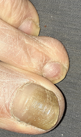
Here are three of my toenails.
Healthy toenail growth is at the base, yet the older section of nail is dark and slightly rough, due to old age -- I had assumed. Though I have suspected years of EMF stress setting up my EMF-damaged toenails for symbiotic microbial scavenging. (Modern Medicine defines this darkening as "fungal infection" and treats it with toxic pharmaceuticals, thereby badly compounding any existing toxic stress.)
For three months now, I've been watching healthy growth at the base of each toenail. This is today's photo.
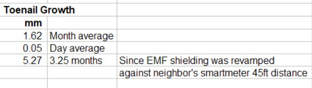
"Tree-Ring" analysis of toenails
Healthy growth and healing should also be occurring throughout my body/mind.
Update 4/26/2022


Fingernails vs Toenails
Generally, "fungal infections" occur more often in toenails than fingernails.
Postulate: Toenail stem cells are more vulnerable to smartmeter radiation because they are already highly EMF stressed. This are nearest to the earth surface where they suffer 24/7 from utility earth current radiation -- more so than any other part of the body. In cities, radiating powerlines are running under the sidewalks.
A Similar Stem Cell Disaster
X-ray photos are another type of EMF, though at high frequency. Decades ago, I had noticed the teeth of a six-month old child who, postnatal, had been carelessly (if not sadistically) exposed to X-rays in a hospital.
At one year, his permanent teeth began to emerge, yet they were were very dull, no sharp edges on the surfaces like normal teeth.
Postulate: The X-ray photos had been taken immediately postnatal, after the child's perfect baby teeth were already formed (though under the gums), and so those baby teeth were perfect. Obviously the permanent teeth stem cells were damaged by those X-rays. Baby teeth and permanent teeth have separate stem cell reservoirs.
Related
1) Smartmeters damage plants
Example 1, Example 2, Example 3
2) Smartmeters damage humans
Example 1, Example 2, Example 3
3) EMF undermines fine muscle coordination
____________________________________________


